

Healio Patellofemoral Update — Simon Talbot, MBChB, FRACS (Orth), Western Health, Melbourne,
Australia
For decades we have assessed the patellofemoral joint by measuring bones. Trochlear dysplasia, patella alta, TT-TG distance and femoral torsion are all measurable, all reproducible, and all embedded in our decision-making. Meanwhile, the structure that actually drives the patella, the quadriceps, has been largely invisible.
This is not for lack of trying. The Q-angle was an attempt to capture the line of force of the extensor mechanism, but it is a two-dimensional measurement taken from bony surface landmarks which ignores the fact that three quarters of the quads attaches to the femur and not the pelvis, and it has never correlated reliably with patellofemoral pathology.
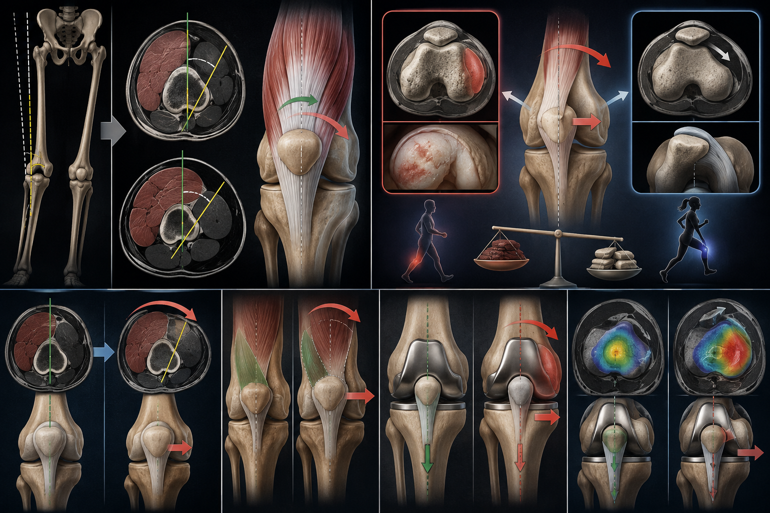
The muscle can now be measured directly. Using standard axial CT or MRI slices, the rotational position of the muscle and the quadriceps tendon relative to the femur can be quantified. A simple measurement we have called the quadriceps tendon axial angle (QTAx) (Figure 1) has excellent correlation with total quadriceps muscle alignment.
What it reveals has changed the way we think about the patellofemoral joint. Quadriceps alignment varies enormously between individuals. In some patients, the entire muscle is rotationally displaced around the femoral shaft — the vastus intermedius migrates laterally, the whole extensor mechanism rotates externally, and the line of force on the patella shifts laterally. Critically, this deformity is completely independent of bony anatomy. In our studies, QTAx shows no correlation with sulcus angle, trochlear inclination, TT-TG, patella height or femoral neck anteversion. It is an isolated soft-tissue deformity.
The first clinical question we asked was whether this deformity is clinically relevant. Isolated lateral facet patellofemoral osteoarthritis (LFPFJOA) is the end-stage of chronic lateral maltracking, so if a lateralised quadriceps force vector is real and clinically relevant, this is where it should show up. In our first comparative study, patients with severe lateral facet patellofemoral osteoarthritis had a mean QTAx of 17.3° external rotation, compared with 3.3° in the control group.
The pattern of the arthritis tells us something important about the mechanism. This is maltracking as a loading problem, not a stability problem. The issue is with the uneven distribution of pressure cross a patella which is constrained within the trochlear groove. So the tracking or direction of movement of the patella can be normal in these patients due to the bony and soft-tissue constraints while the forces acting on the PFJ can be unbalanced. From this study combined with our normal population data we determined that clinically relevant quadriceps alignment occurs in around 15% of the normal population.
The same muscle deformity can also produce instability when combined with inadequate bony and soft-tissue constraints. In our recent study of recurrent patellofemoral instability in native knees mean QTAx in the instability group was 24.8° of external rotation, compared with 2.5° in controls. Using a threshold of 14° (based on our LFPFJOA data) , 90% of instability patients had quadriceps malalignment. On ROC analysis, QTAx was more strongly associated with instability (AUC 0.94) than trochlear dysplasia (0.75) or patella alta (0.78), and remained independently predictive on multivariate analysis.
The unifying concept is imbalance. Rotate the muscle mass externally around the femoral shaft and the force vector on the patella lateralises, regardless of how normal the trochlea is. What happens next depends on the static constraints. If the constraints are inadequate — dysplasia, alta, laxity, the familiar risk factors — the lateralised force overcomes them and the result is recurrent instability. If the constraints hold, the patella never leaves the groove, but it is loaded asymmetrically for decades, and the result is isolated lateral facet osteoarthritis.
In these patients the VMO is not weak or wasted it is rotationally displaced anteriorly so its medialising vector is lost. We have shown that chronic lateral patellar tracking is strongly associated with external rotation of the quadriceps muscle around the femoral shaft, and not with wasting of the vastus medialis. Strengthening a malpositioned muscle cannot restore a force vector. This may explain why some patients fail well-executed rehabilitation.
The consequences follow these patients into arthroplasty. Quadriceps malalignment affects approximately one in five patients undergoing TKA. We have shown that these patients have significantly worse patient-reported outcomes after knee replacement, and that this disadvantage is avoided when the femoral component is externally rotated to compensate for the lateralised force vector. The same imbalance that destroyed the lateral facet in the native knee persists as an unbalanced PFJ after arthroplasty (Figure 2).
I would encourage anyone treating patellofemoral patients to look at the quadriceps alignment. Measuring the quads tendon and muscle, not just the bones, identifies a large group of patients whose lateralising force has a cause we have never previously assessed.
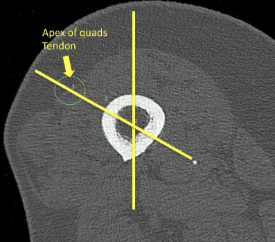
Figure 1. Measurement of the quadriceps tendon axial angle (QTAx) on a standard axial CT slice at the level of the proximal end of the quads tendon. The angle is formed between the anteroposterior axis of the femur (vertical line) and a line from the centre of the femoral shaft to the apex of the quadriceps tendon (arrow). In this patient the QTAx measures 50° of external rotation, indicating severe rotational displacement of the entire quadriceps around the femoral shaft.
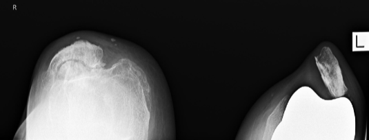
Figure 2. Skyline radiographs of the same patient before and after total knee arthroplasty. Right knee: severe isolated lateral facet patellofemoral osteoarthritis with lateral patellar tilt — the imbalance expressed as chronic lateral facet overload in a native knee whose static constraints held. Left knee: severe lateral patellar tilt and maltracking following TKA due to PFJ imbalance and eventual failure of the medial quadriceps repair.
References
Talbot S, Zordan R, Bennett K, Sasanelli F, Griffith A, Woodford N, Walter WL. Quadriceps tendon malalignment is an independent anatomical deformity which is the primary abnormality associated with lateral facet patellofemoral joint osteoarthritis. Knee Surg Sports Traumatol Arthrosc. 2023;31(12):5950–5961.
Talbot S, Zordan R, Sasanelli F, Sun M. Preoperative quadriceps malalignment is associated with poor outcomes after knee replacement which are avoided by external rotation of the femoral component. Knee Surg Sports Traumatol Arthrosc. 2025;33(4):1418–1427.
Talbot S, Bare J, Zordan R, Suzuki L, Shimmin A. Chronic lateral patella tracking is strongly associated with quadriceps external rotation in relation to the femoral shaft and not with wasting of the vastus medialis. Knee Surg Sports Traumatol Arthrosc. 2025.
Sasanelli F, Zordan R, Suzuki L, Talbot S. Quadriceps malalignment is strongly associated with recurrent patellofemoral instability in native knees. Knee Surg Sports Traumatol Arthrosc. 2026. doi:10.1002/ksa.70448
Further information: www.quadricepsmalalignment.com